

LOOK
Expose the whole forearm & hand.
Look at the:
- Dorsum, Palm,
- Muscles - Thenar, Hypothenar, first dorsal interosseus, ADM, FCU (in forearm)
- Congenital abnormalities
- Open & close hand to quickly assess mass movement of the hand
- - ask for & feel the tender area
- - muscles
- - swellings
- Palmar fascia & 1st web space (for nodules)
- Make a fist (active mass motion)
- Thumb:
- Opposition to all fingers in turn
- Adduction, Abduction, Flexion
- EPL = tested by asking patient to lift thumb up off a table whilst hand held palm down on table
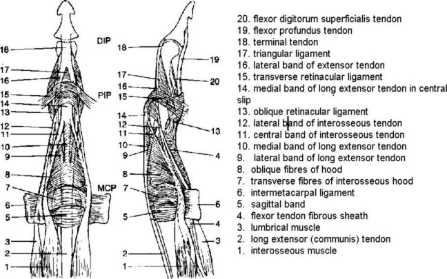
- EDC - extend fingers at MCPJ's
- Interossei - Ask patient to abduct fingers (dorsal interossei); ask patient to adduct fingers (palmar interossei)
- FDS - individually tested by holding other fingers in hyperextension
- FDP - tested by fixing the PIPJ & thus isolating the DIPJ
- Quadriga phenomenon ( a Quadriga = an ancient Greek four horse chariot )
- when testing for FDS the FDP is defunctioned because the FDP tendons are combined, while the FDS muscles are separate in the forearm.
- Following repair or reconstruction of an FDP tendon the tension must be identical to the other FDPs, since the excursion of the combined tendons is equal to the shortest tendon.
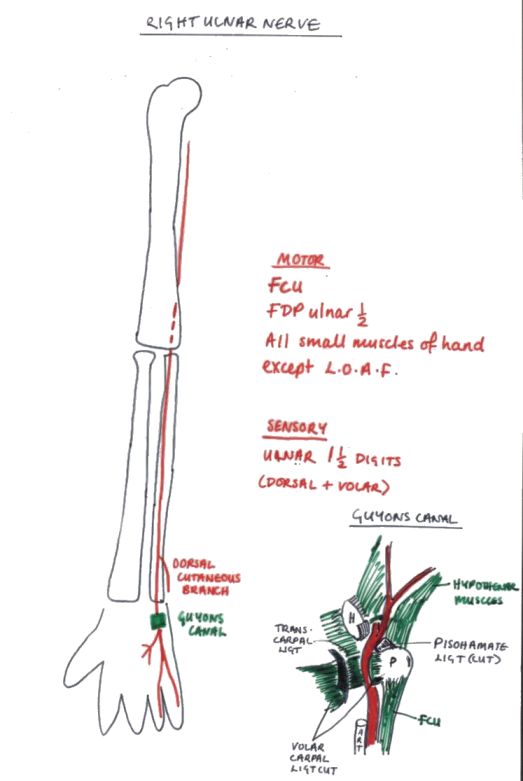
Sensory:
- 1. Autogenous zones:
- Median nerve = volar index finger
- Ulna nerve = volar little finger
- Radial nerve = over 1st dorsal interosseous muscle
- 2. Superficial branch of median nerve - over thenar eminence. Discriminates between a high or low median nerve lesion.
- 3. Dermatomes - C6 = thumb & index finger; C7 = middle finger; C8 = ring & little fingers.
Motor:
Median nerve - test APB with examiners hand over the thenar muscles from the first web space (like shaking hands)
Ulna nerve -
- 1. ADM & 1st dorsal interosseous muscle together, by opening fingers against resistance.
- 2. Testing ADM alone
- 3. Wartenburg's sign - little finger lies abducted due to the unopposed action of EDM.
- 4. Froment's test (Froment described this after watching a train commuter reading his newspaper with on thumb flexed & the other straight)
- Ulna Paradox = less clawing of the fingers than a low lesion, because FDP is involved in high lesions thus flexing MCPJ & relaxing IPJs.
Anterior Interosseous nerve - loss of precise pinch (unable to make 'OK' sign, instead make a square) due to loss of FPL & FDP to index finger.
Posterior Interosseous nerve - Wrist dorsiflexion results in radial deviation (since ECU supplied by PIN, but brachioradialis & ECRL are supplied by the Radial nerve)
Superficial Branch of Radial Nerve:
- Wartenburg's Neuritis (compression at the insertion of Brachioradialis)
- Dellon's sign = active forceful pronation of the forearm & ulnar deviation of the wrist with the elbow extended by the side.
- Tinel's test at the insertion of Brachioradialis
- Power grip / Grasp
- Precision Pinch (AIN)
- Key Grip Pinch
- Strength - tested on Dynamometer or Sphygmanometer
Allens test - Ask patient to clench fist; compress both the radial & ulna arteries together with thumbs; Patient relaxes hand; Release one artery & observe capillary refill
1. Differentiate Intrinsic contracture from forearm flexor contracture
Flexing the wrist relaxes the FDS & FDP (long flexor) tendons; if patient can then flex the IPJ's with the wrist flexed there is intrinsic tightness, if they cannot it is a Volkmann's contracture.
2. Bunnel-Littler Test
For intrinsic tightness.
1) With the MCPJ in extension the intrinsics are put on a stretch. Try to flex the PIPJ with MCPJ in extension.
If it doesn't flex = tight intrinsics or joint capsule contracture.
2) With MCPJ in flexion the intrinsics are relaxed. Thus if unable to flex PIPJ= tight capsule.
NB- prior to test check that passive motion of PIPJ is possible (i.e. normal PIPJ)
Tight intrinsics occur in: 'Intrinsic Plus' hands due to ischaemia or fibrosis of intrinsics or RA.
3. Differentiate a Lumbrical Plus Finger from an Intrinsic Plus Finger:
Lumbrical Plus Finger is manifested by intrinsic plus attitude in involved finger on attempted flexion: ( with MCPJ flexion there will be IP extension ); FDP becomes an extensor of the PIP joint; when FDP relaxes FDS can work with less antagonism and PIP can flex; treatment may involve division of the lumbrical; Causes: (lumbrical tighter than FDP) - FDP laceration or rupture distal to the Lumbrical Origin from FDP (the proximal end of the lacerated FDP tendon will retract proximally, drawing the attached lumbrical proximally as well. The effect is increased tension on the radial lateral band, which causes the PIP joint to extend); 2) Amputation of the Distal Phalanx (distal to central slip insertion); 3) Excessively Long Tendon Graft.
Bouvier's Test
- To determine if PIPJ capsule & ext. mech. are working normally.
- If PIPJ capsule & ext. mech are functioning normally then blocking MCPJ hyperextension allows IPJ extension.
- Positive test occurs as a result of: attenuation of central slip, adherent central slip at PIPJ or volar subluxation of lateral bands.
Tests for Traumatic Bouttoniere Deformity
Elson's Test = Put finger over edge of table, with PIPJ flexed to 90deg. & ask Pt to extend against resistance. Weakness of resisted extension of PIPJ & hyperextension of DIPJ occurs if the central slip is ruptured. More Detail - Original Article
Passive test = flex wrist & MCPJs. Poor passive resistance to pushing over middle phalanx indicates weak extensor mechanism.
Boye's Test (1970) - If the PIPJ is held passively extended, it is then possible for the normal individual to flex the terminal interphalangeal joint in isolation. However, if the central slip has been ruptured, there is increasing difficulty in performing this action. Unfortunately this test only becomes positive when the proximal part of the ruptured central slip has retracted and become adherent to the surrounding tissues.
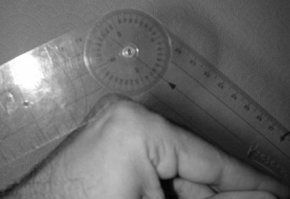
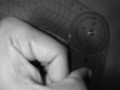
How to measure fixed contractures in the MCPJs and PIPJs in dupuytrens etc.
- Pronate the hand so that the dorsum faces you.
- Then keep a goniometer on the dorsum of the MCPJ.
- For assessing the PIPJ flex the MCPJ as much as possible - this reduces the possible chance for a fixed contracture of the MCPJ to contribute to a contracture of the PIPJ
Compartment | Contents | Pathologic Conditions |
1 | APL & EPB | DeQuervain's Disease |
2 | ECRL & ECRB | Tennis elbow |
3 | EPL | Rupture at Lister's tubercle |
4 | EDC & EIP | Extensor Tenosynovitis |
5 | EDM | Rupture (rheumatoid) |
6 | ECU | Snapping at ulnar styloid |
Tidak ada komentar:
Posting Komentar